
Magnetic Resonance Imaging (MRI) is a non-invasive procedure in which the combination of radio waves – Read more
Computerized Axial Tomography, frequently abbreviated simply as CAT or CT scanning uses special equipment – Read more
Stay right here on the Peninsula to take advantage of our world class Women’s Imaging Services starting with – Read more
Ultrasound or sonography is a non-invasive way to “see” inside the body using sound waves – the principles are – Read more
Nuclear medicine uses a small amount of radioactive material or tracer, a special gamma camera – Read more
Bone Densitometry has become the gold standard for measuring the density of your bones. Sometimes referred – Read more
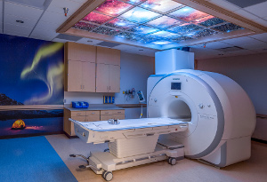
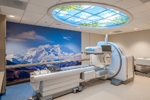
Magnetic Resonance Imaging
Magnetic Resonance Imaging (MRI) is a non-invasive procedure in which the combination of radio waves and a powerful magnet linked to a computer are used to create remarkably clear and detailed images of internal organs and tissues without the use of radiation. Each MRI examination produces hundreds of pictures from a variety of different angles. One way to think of MRI 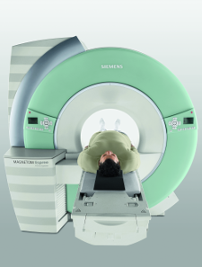 is as you would a loaf of bread and the many slices in it. These “slices” show the difference between normal and abnormal tissue making MRI very useful in diagnosing abnormalities. MRI has proven very valuable for the diagnosis of many conditions in the following areas of your body:
- Brain
- Spine
- Abdomen and pelvis
- Vascular structures
- Joints such as the knee and shoulder
- Muscles, cartilage, tendons and ligaments
How to prepare for your MRI examination:
Unless you’re told otherwise, you may follow your regular daily routine and take food and medications as usual. We’re necessarily vigilant about your safety while in the strong magnetic field of our scanner. Prior to your scan we’ll ask you to complete a safety questionnaire to ensure you’re a good candidate for an MRI scan. You’re encouraged to check with our registered technologist if you have any questions or concerns about any implanted object or health condition that could affect your MRI. Be sure to let us know if you have a:
- Pacemaker (unless it’s one of the new MRI compatible types!)
- Aneurysm clips
- History of working with metal
- Implanted infusion device
- Inner ear implant
- History of bullet or shrapnel injury
- Metallic plates, pins, screws
- IUD
- Tattoos or permanent makeup
- Are pregnant
What to expect during your MRI examination:
You’ll be asked to change into a pair of scrub pants and a top for your scan. We’ll secure your valuables outside the scanning suite since you’ll not be able to bring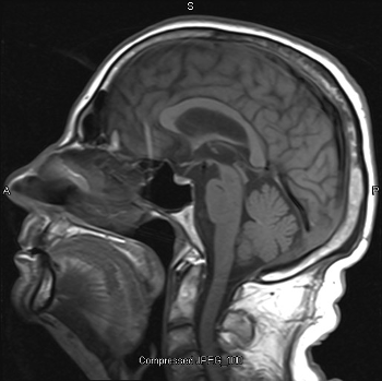 them into the room with you. Your registered technologist will make you as comfortable as possible on a table that moves into the MRI machine during the scan. The area of interest needs to be in the center of the magnet, which is approximately six feet deep overall. A special coil may be placed over the area to be examined to improve the quality of the images. It’s very important that you remain as still as possible during each scanning sequence to allow us to obtain the best images for your exam. You’ll usually be alone in the exam room during the MRI procedure however, your technologist will be able to see, hear and speak with you at all times using a two-way intercom. S/he will update you on the progress and duration of your scan. We’ll provide either ear plugs and/or headphones for you to listen to music during your exam. We also have the ability to plug your personal iPod or MP3 player into our sound system. During scanning you’ll hear noises ranging from grating to thumping to clicking or tapping sounds as the scanner changes to different sequences or “pictures”. It’s normal for the area of your body being imaged to feel slightly warm – let your technologist know if this bothers you. For some MRI studies, a contrast agent called gadolinium (not iodine) is injected through an IV and may be used to improve the visibility of certain tissues or blood vessels. Please let your technologist know if you have any history of kidney problems. Plan to be a guest in our Imaging Department for about an hour for your MRI study.
After your MRI examination:
You should not feel any after effects from your MRI exam and can immediately resume your normal daily routine. Your technologist will send your images to a Board Certified Radiologist with special training interpreting MRI examinations. S/he will analyze the images and send the results to your healthcare provider. Your provider will discuss the results with you. New technology allows us to distribute reports and images over the Internet to many providers and facilities.
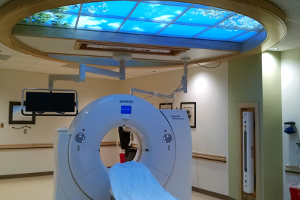
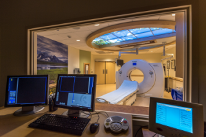
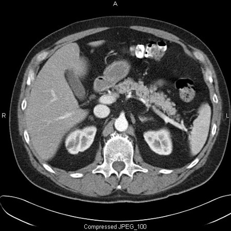
Computed Tomography
Computerized Axial Tomography, frequently abbreviated simply as CAT or CT scanning uses special equipment to transmit a fan-shaped x-ray beam as it rotates 360 degrees around your body. Across from the x-ray beam, detectors “read” the amount of x-ray that passes through your body. A digital computer together with the rotating x-ray device and detectors is used to create detailed cross sectional, multi-planar or 3 dimensional images or “slices” of your tissues and organs. Imagine the body as a loaf of bread and you’re looking at one end of the loaf. As you remove each slice of bread, you can see the entire surface of that slice from the crust to the center. Be assured that during each CT scan, our staff practice the principles of ALARA which stand for As Low As Reasonably Achievable – we use the lowest dose of radiation in achieving the best image quality in making a diagnosis. The body is seen on CT scan slices in a similar fashion from the skin to the central part of the body part being examined. When these views are added together, a three-dimensional picture can be obtained. Different body parts absorb the x-rays to varying degrees. CT takes advantage of the different absorption characteristics of tissues in your body to show anatomical images with great clarity. Bone absorbs the most and your lung the least amount of x-ray. Unlike MRI, CT exams can be performed even if you have a pacemaker or other implanted medical device. CT is commonly used to image the:
- Lungs

- Abdomen
- Brain
- Soft tissues
- Blood vessels
- Spine
- Skeletal structures
- Guidance for biopsy procedures
- Plan surgery and different treatments
How to prepare for your CT examination:
Depending on the part of the body being scanned you may be given special instructions regarding eating and drinking prior to your procedure. If we’re scanning your abdomen you may be given oral contrast to start drinking the night before your examination. The oral contrast helps highlight your gastrointestinal tract so the radiologist can clearly differentiate tissues. You should wear comfortable, loose-fitting clothing for your CT scan and remove any metal objects that might interfere with the image results. Alternatively you may be asked to change into a hospital gown for your examination. Plan to be a guest in our Imaging Department for about 45 minutes for your CT scan. Let your registered technologist know if you:
- Have a known allergy to IV iodine contrast

- Have heart failure
- Have diabetes and are on medications
- Have a history of kidney problems
- Are pregnant or might be
What to expect during your CT examination:
A CT scanner looks like a large donut with a narrow table in the middle. The technologist begins by positioning you on the CT table as comfortably as they can so you’re able to hold still during the study. Your body may be supported by pillows or bolsters to help you remain in the proper position during the scan. Once the exam begins, the table will move slowly through the round opening of the CT scanner while the x-ray tube inside the “donut” rotates or spirals continuously around your body. You may hear a clicking or buzzing sound as the table moves through the machine. Due to the speed of spiral CT, we’re usually able to scan your entire body during a single breath hold. For most exams you’ll be alone in the room during the scan but the technologist will direct and monitor your exam from an adjacent room and can see and hear you at all times. Certain exams require an IV injection of iodine contrast material to highlight certain anatomy and blood vessels. The injection may give you a warm sensation, a metallic taste in your mouth or make you feel as though you’re urinating (but you’re not!) If your experience any of these sensations please be assured that they only last for a few seconds.
After your CT examination:
You should not feel any significant effects from your CT exam and can immediately resume your normal daily routine. If you drank oral contrast for your study you may experience some diarrhea. If we gave you an IV injection your technologist will encourage you to drink extra fluids to help prevent the iodine/salt solution from dehydrating you. Your technologist will send your images to a Board Certified Radiologist with special training interpreting CT examinations. S/he will analyze the images and send the results to your healthcare provider. Your provider will discuss the results with you. New technology allows us to distribute reports and images over the Internet to many providers and facilities.
3D Digital Breast Tomosysnthesis
We’re excited to let you know we’re now offering an advanced form of mammography called 3D or Digital Breast Tomosynthesis (DBT) With DBT, the x-ray tube moves in an arc over the compressed breast capturing multiple images of each breast from different angles. These digital images are then reconstructed or “synthesized” into a set of three-dimensional images by a computer. These three dimensional image sets help minimize the tissue overlap that can hide cancers or make it difficult to distinguish normal overlapping tissue from tumors. 3D Mammography has the ability to detect an average of 41% more invasive breast cancers and also reduce call back rates by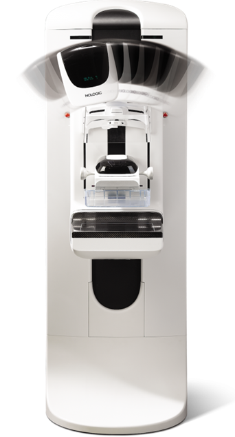 15%. Additionally, the radiation dose from the 3D mammogram is only ever so slightly more than a 2D digital mammogram. Every two minutes there’s a new breast cancer diagnosis made. Most medical experts agree that successful treatment of breast cancer is often linked to early diagnosis. 3D Mammography plays a critical role in early detection of breast cancer because it can show changes in the breast up to two years before you or your physician can feel them. For your mammogram we’ll ask you to change into a gown. If you’re wearing any powder, deodorant, lotion or perfume on your breast or under arm area we’ll ask you to thoroughly wash these products off – if left on your skin they can show up on the mammogram mimicking an abnormality. During mammography your breast is placed on a cassette, compressed with a paddle and exposed to a small dose of radiation to produce an image. Compression of your breast is necessary to allow for minimal x-ray dosage and maximum tissue visualization. If you experience breast tenderness as part of your monthly cycle it may be best to schedule your appointment before or after that time to avoid any discomfort from the mild compression we use. As with all radiologic exams be sure to let your technologist know, before your exam, if there’s any chance you might be pregnant. Occasionally the initial mammographic images don’t provide enough information to determine whether something is normal or abnormal. If a finding or spot seems suspicious, the radiologist may recommend additional views or further diagnostic studies.
- Breast Imaging Services we offer:
- Screening mammography – no order from your provider needed, just their name to send results to. The American Cancer Society recommends screening mammography every year for women beginning at age 40.
- Diagnostic mammography – a problem solving exam (when a lump or other abnormality needs to be evaluated) directed by our on-site radiologist
- Computer Aided Detection (CAD) – a computer program that uses pattern recognition to assist the radiologist in looking for breast abnormalities on your mammogram
- Breast MRI with CAD – another modality used in problem solving of questionable mammogram results
- Stereotactic and ultrasound guided biopsies – tissue sampling without having to undergo a surgical procedure
- Ductography – to evaluate the source of abnormal nipple discharge
- Needle localization – for surgical planning
Spend 15 minutes getting your mammogram to help guard against this serious disease. Digital Breast Tomosynthesis mammography is now available at Central Peninsula Hospital. If you had a mammogram at another facility be sure to let us know so we can get to work obtaining your prior exams. The radiologist will look closely at all your studies to uncover any small changes that have occurred from year to year and send the results to your provider. We’ll also mail (snail or -email) you a letter to let you know whether your exam is normal or requires additional imaging.
Schedule your mammogram today: (907) 714-4542
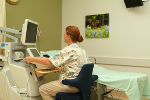
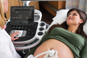
Ultrasound Services
Ultrasound or sonography is a non-invasive way to “see” inside the body using sound waves – the principles are similar to sonar used by boats and submarines. When a sound wave strikes an object, it bounces back, or “echoes”. By measuring these echo waves, it is possible to determine how far away the object is and its size, shape and whether the object is solid, filled with fluid or both. During the procedure the sonographer presses a transducer (a small, smooth plastic device about the size of a deck of cards) against your skin that both sends the sound wave and “listens” for echoes returning from tissues in your body. These unique waves are instantly measured and displayed by a computer, which in turn creates a real-time picture on the monitor. Several frames of the pictures are typically captured as still images. Small loops or “movies” of “real-time” images also may be saved. Common uses of ultrasound include:
Obstetrical

Gynecological/pelvic
Abdominal
Vascular
Heart
Breast
Thyroid
Scrotal
Biopsy procedures
How to prepare for your ultrasound examination:
You should wear comfortable, loose-fitting clothing for your ultrasound exam. You may be asked to change into a gown for your procedure. Other preparation depends on the type of examination you will have. For some scans you may be instructed not to eat or drink for as many as 12 hours before your appointment. For others you may be asked to drink up to six glasses of water two hours prior to your exam and avoid urinating so that your bladder is full when the scan begins. Most medications are okay to take with a sip of water. To help us obtain the best diagnostic exam for you it’s important to follow any preparation instructions given.
What to expect during your ultrasound examination:
For most ultrasound exams you will be positioned lying on your back on a padded exam table. You may be asked to move into different positions during the procedure or hold your breath for short periods of time. A clear, warm, water-based gel is applied to the area of the body being studied to help the transducer make secure contact with the body and eliminate air pockets between the transducer and the skin that can block the sound waves from passing into your body. A Registered Diagnostic Medical Sonographer will press the transducer firmly against your skin in various locations and at various angles to carefully look at the area of interest and obtain the desired images. If scanning is performed over an area of tenderness you may feel some pressure – especially if we have to scan over a sore spot. Be sure to let your sonographer know if this pressure is too uncomfortable. Plan to be a guest in our Imaging Department for 30 – 60 minutes to complete your ultrasound examination.
After your ultrasound examination:
The gel used during the procedure is not harmful to your skin or clothing. You should not feel any after effects from your ultrasound exam. Your sonographer will send your ultrasound images to a Board Certified Radiologist with special training interpreting diagnostic examinations. S/he will analyze the images and send the results to your healthcare provider. Your provider will discuss the results with you. New technology allows us to distribute reports and images over the Internet to many providers and facilities.
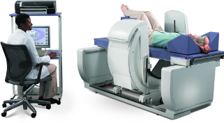
Nuclear Medicine
Nuclear medicine uses a small amount of radioactive material or tracer, a special gamma camera (like a big Geiger counter) and a computer to form images. The tracer is commonly injected, but may also be swallowed or inhaled. You will need to wait a few minutes, hours or even a few days before having your scan. This allows for the tracer to concentrate in the part of your body being studied. One of the unique features of a nuclear medicine scan is that, based on the degree to which the tracer is absorbed or “taken up” by a particular organ, the examination shows the “function” as well as the size, shape and position of the organ or tissue being evaluated as opposed to just a picture. This helps to determine if the organ is working properly. Commonly performed nuclear medicine exams involve imaging of the following:

Heart – to identify blood flow to the heart or a specific vessel. We also scan to evaluate the extent of damage to the heart muscle after a heart attack.
Bones – to evaluate degenerative or arthritic changes in joints, bone disease or the cause of bone pain or inflammation
Gallbladder – identify inflammation or abnormal function
Thyroid – measures function of the gland. Nuclear Medicine also offers treatment options for certain thyroid conditions.
Lung – used to locate a blood clot or embolism in the lung
Stomach – how it empties
How to prepare for your nuclear medicine scan:
It’s very important that you follow any preparation instructions we provide for your examination. These may include specific information about eating and whether or not you should take your routine medications. Wear loose comfortable clothing for your test. Be sure to let your technologist know about all medications and herbs you’re currently taking. Also, be sure to mention any recent CT imaging studies involving an iodine injection or oral contrast media taken such as for an Upper GI or Barium Enema study. If there’s any chance you might be pregnant or are breast feeding let your technologist know prior to your radiotracer injection.
What to expect during your nuclear medicine examination:
Your examination will be performed by a registered nuclear medicine technologist. It’s possible that you will come in first for the administration of the tracer and then return later for the actual scan. Sometimes the entire procedure may be done during one visit. There are also exams that require multiple visits in a day or over a few days. The actual imaging time varies anywhere from less than an hour to several hours. When the exam starts you’ll lie on a special exam table and be made as comfortable as possible. You’ll be able to listen to what ever type of music you like during most nuclear medicine procedures. It’s important for you to be able to lie still for the study. The technologist will be with you during the exam to assist in making you comfortable. The gamma camera may be close to the area of your body that’s being examined while the images are formed. Before you leave our department the technologist will review your images to ensure that we’ve obtained all the information our radiologist needs to interpret your study.
After your nuclear medicine examination:
You may resume your normal activities after your scan. If any special instructions are necessary you’ll be informed by your technologist. Through the natural process of radioactive decay the small amount of radiotracer in your body will lose radioactivity over time. It will also pass out of you through your urine or stool within 48 to 72 hours. You should also drink plenty of water to help flush the radioactive material out of your body as instructed by your technologist. Your technologist will send your images to a Board Certified Radiologist with special training interpreting nuclear medicine examinations. S/he will analyze the images and send the results to your healthcare provider. Your provider will discuss the results with you. New technology allows us to distribute reports and images over the Internet to many providers and facilities.
Bone Densitometry
Bone Densitometry has become the gold standard for measuring the density of your bones. Sometimes referred to as DEXA (dual energy x-ray absorbtiometry) scan, bone densitometry is the test for the evaluation of osteoporosis (the loss of bone minerals.) The results of your exam are compared to others whose age, sex and ethnic background are similar to yours. The measurement of bone mineral is very closely related to bone strength and your potential for bone fracture. As men and women age, their risk factors for osteoporosis increase. Common risk factors include:
- Postmenopausal, including early or surgical

- Age
- Previous bone fracture
- Family history of osteoporosis
- Certain medications
- Caucasian or Asian decent
- Thin or small build
- Eating disorder
- Smoking
- Alcohol abuse
- Inactive lifestyle
- Low calcium intake
- Vitamin B deficiency
DEXA uses low-dose x-ray that checks for signs of mineral loss and bone thinning. We commonly scan your hip, spine and/or wrist for this exam. DEXA is a simple and painless test that takes about 15 minutes while you lie comfortably on our table. Metal buttons, buckles or zippers interfere with the test so you may be asked to change into a gown. Our radiologist will interpret the results of your scan and send the results to your provider. S/he will discuss the results with you. After an initial DEXA scan is done, subsequent tests are useful to monitor your rate or response to therapy to slow bone loss.
Our DEXA system at CPH also has the ability to perform an accurate assessment for Body Composition Analysis (BCA.) These scans are performed in a fashion similar to a DEXA study. For more than three decades the prevalence of obesity has increased rapidly among both adults and children. Today, more than two thirds of U.S. adults are considered overweight or obese and more than 97 million Americans are affected by obesity related illnesses. The demand for accurate body composition analysis is growing as health care providers realize its value in identifying serious health risks that result from obesity including heart disease and type 2 diabetes. The BCA scan produces color images displaying the distribution of fat, lean tissue and bone and translates the information into a report that lets you know exactly how you’re progressing towards your weight management goal. You do not need a physician’s order for the BCA evaluation.