For COVID information please click the following link  >>More Info

Ultrasound

Ultrasound Services

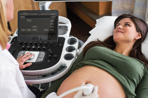
Ultrasound or sonography is a non-invasive way to “see” inside the body using sound waves – the principles are similar to sonar used by boats and submarines. When a sound wave strikes an object, it bounces back, or “echoes”. By measuring these echo waves, it is possible to determine how far away the object is and its size, shape and whether the object is solid, filled with fluid or both. During the procedure the sonographer presses a transducer (a small, smooth plastic device about the size of a deck of cards) against your skin that both sends the sound wave and “listens” for echoes returning from tissues in your body. These unique waves are instantly measured and displayed by a computer, which in turn creates a real-time picture on the monitor. Several frames of the pictures are typically captured as still images. Small loops or “movies” of “real-time” images also may be saved. Common uses of ultrasound include:

  • Obstetricalultrasound.png

  • Gynecological/pelvic

  • Abdominal

  • Vascular

  • Heart

  • Breast

  • Thyroid

  • Scrotal

  • Biopsy procedures

How to prepare for your ultrasound examination:

You should wear comfortable, loose-fitting clothing for your ultrasound exam. You may be asked to change into a gown for your procedure. Other preparation depends on the type of examination you will have. For some scans you may be instructed not to eat or drink for as many as 12 hours before your appointment. For others you may be asked to drink up to six glasses of water two hours prior to your exam and avoid urinating so that your bladder is full when the scan begins. Most medications are okay to take with a sip of water. To help us obtain the best diagnostic exam for you it’s important to follow any preparation instructions given.

What to expect during your ultrasound examination:

For most ultrasound exams you will be positioned lying on your back on a padded exam table. You may be asked to move into different positions during the procedure or hold your breath for short periods of time. A clear, warm, water-based gel is applied to the area of the body being studied to help the transducer make secure contact with the body and eliminate air pockets between the transducer and the skin that can block the sound waves from passing into your body. A Registered Diagnostic Medical Sonographer will press the transducer firmly against your skin in various locations and at various angles to carefully look at the area of interest and obtain the desired images. If scanning is performed over an area of tenderness you may feel some pressure – especially if we have to scan over a sore spot. Be sure to let your sonographer know if this pressure is too uncomfortable. Plan to be a guest in our Imaging Department for 30 – 60 minutes to complete your ultrasound examination.

After your ultrasound examination:

The gel used during the procedure is not harmful to your skin or clothing. You should not feel any after effects from your ultrasound exam. Your sonographer will send your ultrasound images to a Board Certified Radiologist with special training interpreting diagnostic examinations. S/he will analyze the images and send the results to your healthcare provider. Your provider will discuss the results with you.  New technology allows us to distribute reports and images over the Internet to many providers and facilities.

Email to a Friend

Share this with friends!

Email
Facebook
Twitter
Pinterest
LinkedIn
LinkedIn
Digg
StumbleUpon
Reddit
Tumblr
VK
OK
Skype
Mix
Telegram
Pocket
XING
WhatsApp